// NATURE NEWS — SPAZIO & SCIENZA
The lipidomic architecture of the mouse brain
Nature
(2026) Cite this article
Lipids are fundamental components of the brain, crucial for synaptic transmission and signal propagation. Altered brain lipid composition is associated with common and rare neuropathologies, yet the spatial organization of the mammalian brain lipidome remains insufficiently characterized compared with other modalities1,2,3,4,5,6. Here we mapped the membrane-lipid architecture of the adult mouse brain at micrometric scale, across sexes and during pregnancy. This lipid brain atlas reveals that lipids describe a fine-grained biochemical structure that aligns with functional anatomy. Membrane-lipid spatial heterogeneity clusters into territories, which we termed ‘lipizones’. Lipizones partially mirror cell-type territories, but also capture distal axon terminals. Through lipizones, we (1) reveal the organizing principles of the grey matter lipidome, related to connectivity and cytoarchitecture; (2) discover a new axis of oligodendrocyte heterogeneity in the white matter; and (3) find biochemical zonation in the choroid plexus and in the ventricular walls. We show that this lipidomic architecture can adapt to changing physiological needs. In the brain of pregnant female mice, the white matter is metabolically activated and the cortex undergoes a lipizone-specific remodelling that is particularly pronounced in layer 4. These results are a foundational resource (https://lbae-v2.epfl.ch/) poised to reshape the understanding of lipids in brain development, physiology and pathology.
Lipids account for most of the brain dry weight7. They form the cell membranes that constitute myelin, axons, dendrites and synapses. Seminal studies have examined the lipid composition of the mammalian brain across anatomical structures and cell classes8,9,10,11,12. However, a systematic survey of the brain lipid metabolic architecture13 in relation to cell-type composition, functional anatomy, connectivity and physiological variation is lacking.
To fill this gap, we used matrix-assisted laser desorption/ionization mass spectrometry imaging (MALDI–MSI), previously used to map brain lipids in two-dimensional (2D) and three-dimensional (3D) imaging11,12,14. We mapped the distribution of 172 lipids across 109 brain sections from 11 mice, covering the entire brain volume (5 µm laser spot size and 25 µm interpixel distance, thus sampling portions of one to four cells per pixel). We identified 539 lipidome-defined brain clusters, which we termed ‘lipizones’. We characterized lipizones in relation to anatomy, cell-type composition, cell compartments, connectivity and biochemistry. We investigated inter-individual and sex-related differences and, to probe physiological variation, charted the spatial lipidome in pregnant mice.
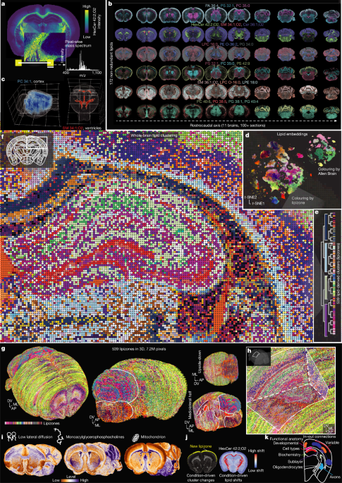
We built a comprehensive lipidomic atlas of the adult mouse brain, measuring coronal sections from male and female brains. Using MALDI–MSI in positive ion mode and the unified Mass Imaging Analyser (uMAIA)15, we mapped 26,874 peaks across 7.2 million pixels (Fig. 1a,b and Supplementary Fig. 1a–o; Methods), which were warped into the Allen Brain Atlas (ABA) Common Coordinate Framework (CCF) (ABA CCFv.3 (ref. 16)) for systematic comparison with anatomy and other modalities (Supplementary Fig. 1b–d; Methods). A total of 1,400 peaks passed noise quality control (Supplementary Fig. 1e–j and Supplementary Tables 1 and 2; Methods).
a, Schematic of a MALDI–MSI experiment measuring the mass spectrum of individual 5 µm desorption points (pixels), 25 µm interpixel distance, along an adult brain coronal section; HexCer 42:2;O2 distribution is displayed as an example. b, Overview of MALDI–MSI composite images for selected annotated lipids along the rostrocaudal axis. c, Three-dimensional distributions of selected lipids obtained interpolating serial sections. d, t-Distributed stochastic neighbour embedding (t-SNE) of pixels in lipid space, coloured by lipizone and by Allen Brain region. e, T