// NATURE NEWS — SPAZIO & SCIENZA
The Virtual Tissues foundation model resolves spatial proteomics across scales
Nature
(2026) Cite this article
Spatial proteomics technologies have transformed our understanding of complex tissue architecture in cancer but present unique challenges for computational analysis1. Each study uses a different marker panel and protocol, and most methods are tailored to single cohorts, which limits knowledge transfer and robust biomarker discovery. Here we present Virtual Tissues (VirTues), a general-purpose foundation model for spatial proteomics that learns marker-aware, multi-scale representations of proteins, cells, niches and tissues directly from multiplex imaging data. From a single pretrained backbone, VirTues supports marker reconstruction, cell segmentation and typing, niche annotation, spatial biomarker discovery and patient stratification, including zero-shot annotation across heterogeneous panels and datasets. In triple-negative breast cancer, VirTues-derived biomarkers predict anti-PD-L1 chemo-immunotherapy response2 and stratify disease-free survival in an independent cohort3, outperforming state-of-the-art biomarkers derived from the same datasets and current clinical stratification schemes.
Tissues, particularly in cancer, display pronounced heterogeneity across patients, disease stages and within individual tumours, evident in diverse cell phenotypes, states and spatial organization1. Tumour development and therapy response depend not only on cancer cells but also on their interactions with the surrounding environment, making the spatial organization and function of the tumour microenvironment (TME) central to cancer treatment.
Capturing this organization requires molecular imaging beyond traditional methods4. Spatial proteomics technologies, including multiplexed immunofluorescence and imaging mass cytometry (IMC)5, now measure dozens to hundreds of proteins in intact tissue at subcellular resolution, revealing tumour-immune interactions, prognostic spatial niches and mechanisms of therapy resistance across cancer types6 (Fig. 1a).
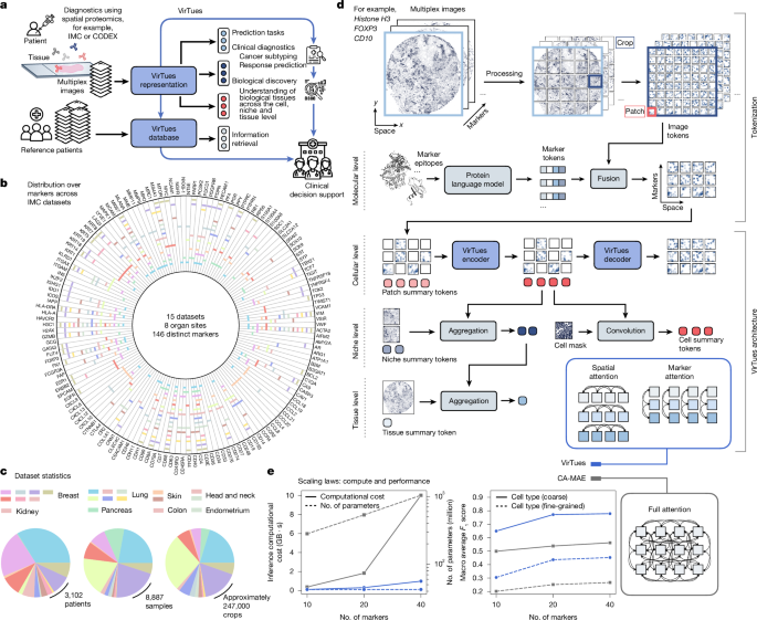
a, Flow chart depicting VirTues capabilities. VirTues converts highly multiplexed images of tissue to virtual tissue representations useful for clinical and biological investigations at cell, niche and sample level, including the retrieval of similar tissue samples for clinical decision support. b, VirTues is trained and evaluated on 15 IMC datasets with a focus on tumours and their TMEs originating from 8 different organ sites, measuring 146 distinct markers in total. The polar plot depicts used marker panels per dataset. A legend of the dataset colour codes is provided in Extended Data Fig. 1a. c, Origins and sizes of datasets in terms of patients, tissue samples and 256 × 256 image crops. d, Multiplexed images are processed crop-wise into three-dimensional grids of image tokens, representing patches of each marker at each position. Marker tokens, derived from a PLM, are fused with the respective image tokens using a linear projection and addition. VirTues is a new ViT architecture trained with a masked autoencoding objective. Input tokens are concatenated with patch summary tokens, which are initialized with learnable weights. During inference, VirTues’ encoder processes this set of tokens. The encoded patch summaries are subsequently convolved with the cell segmentation mask into cell summary tokens or aggregated to niche and tissue summary tokens. For training, a random subset of tokens is selected and masked independently for each channel. VirTues’ decoder predicts channel-wise reconstructions receiving as input the encoded, non-masked tokens from the target channel along with all patch summary tokens. VirTues encoder uses sparse attention mechanisms restricting direct token interactions to either positions (marker attention) or channels (spatial attention). e, Comparison of computational cost (left) and prediction performance (right) between CA-MAE31 and VirTues as a function of the number of markers used.
Yet the flexibilit