// NATURE NEWS — SPAZIO & SCIENZA
Mitochondrial fission factor senses and governs ferroptosis
Nature
(2026) Cite this article
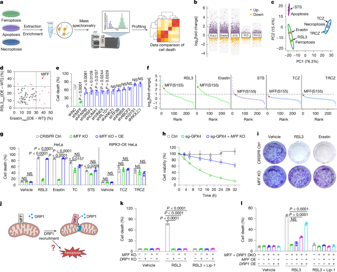
Ferroptosis is a form of regulated cell death driven by iron-dependent lipid peroxidation and is implicated in diverse pathological states1,2,3,4. Although mitochondria and other organelles are increasingly being recognized as important modulators of ferroptosis5,6,7, a unifying mechanism that couples organelle dynamics to ferroptotic execution has remained elusive. Here using quantitative phosphoproteomics, we identified mitochondrial fission factor (MFF) as a key ferroptosis-selective regulator. Mechanistically, the lipid mediator 17-HETE promotes phosphorylation of MFF at Ser155, which triggers the coordinated fragmentation and dysfunction of mitochondria and peroxisomes. This remodelling intensifies interorganelle crosstalk, amplifies oxidative stress and accelerates ferroptotic death. To monitor this phosphorylation event in living cells, we developed MFF–SPARK, a phase-separation-based biosensor, for real-time tracking of ferroptosis through MFF activation. Using MFF–SPARK, we identified PKCβ and DUSP22 as a coordinated kinase–phosphatase pair that governs MFF phosphorylation. We also discovered avermectin B1 as a pharmacological activator of the PKCβ–MFF axis, which can sensitize tumours to ferroptosis in vivo. Together, our findings establish MFF phosphorylation as a central regulatory node in ferroptosis-associated organelle remodelling and provide a conceptual framework and toolbox for monitoring and pharmacologically interrogating ferroptosis.
This is a preview of subscription content, access via your institution
Access Nature and 54 other Nature Portfolio journals
Get Nature+, our best-value online-access subscription
Prices may be subject to local taxes which are calculated during checkout
All data supporting this study are available in the article, its Supplementary Information and deposited databases. The phosphoproteomic data have been deposited into ProteomeXchange via iProX (accession PXD079759). The metabolomic data have been deposited to Mendeley Data (https://doi.org/10.17632/93m6cpvhcp.1). Public databases referenced in this study (KEGG, HMDB, LIPID Maps and Gene Ontology) are described in the Methods. Gene OE screen data for Fig. 1d are provided in Supplementary Table 1. Uncropped western blot scans are provided in Supplementary Fig. 1. FACS gating strategies are provided in Supplementary Fig. 2. Source data are provided with this paper.
No custom code was used in this study. All data analyses were performed using publicly or commercially available software as detailed in the Methods.
Dixon, S. J. et al. Ferroptosis: an iron-dependent form of nonapoptotic cell death. Cell 149, 1060–1072 (2012).
Article
CAS
PubMed
PubMed Central
Google Scholar