// NATURE NEWS — SPAZIO & SCIENZA
Sexual dimorphism in epidermal stem cell dynamics
Nature
(2026) Cite this article
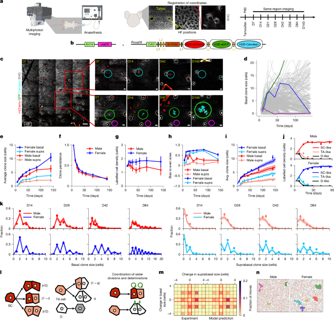
The epidermis is a stratified epithelium comprising proliferative basal cells and differentiated suprabasal cells that are continuously renewed throughout life. Conflicting theories have been proposed regarding the heterogeneity of the basal cell layer, with some studies proposing that all proliferative cells have self-renewal potential during homeostasis1,2,3,4,5. Meanwhile, others posit a model in which stem cell differentiation occurs through an intermediate transit-amplifying-like progenitor state with strictly limited proliferative potential6,7,8,9,10. Here, to determine the heterogeneity and pattern of cell fate, we performed intravital time-lapse imaging to track the fate of lineage-labelled cells in the skin interfollicular epidermis. Results from quantitative analyses of clonal dynamics support a two-compartment model of epidermal homeostasis, whereby basal cells are organized in a stem cell–transit-amplifying cell hierarchy. Critical to these conclusions was the discovery of sexual dimorphism in epidermal fate, with female mice showing a highly synchronized pattern of cell division, differentiation and delamination that contributes to accelerated wound healing. Single-cell RNA sequencing, transcriptional profiling and clonal analyses of castrated mice revealed that male sex hormones promote stem cell differentiation and delamination. In summary, our study provides insight into the functional identity of the stem cell population and identifies sex-specific differences in the regulation of epidermal stem cells and transit-amplifying cell fate.
The skin epidermis is a compartmentalized tissue that is subdivided into the hair follicle, interfollicular epidermis (IFE), infundibulum and sebaceous gland. All of these cell types are maintained by distinct populations of stem cells (SCs)11,12. The IFE is a stratified epithelium comprising a proliferative basal layer and multiple layers of differentiated suprabasal cells11. Following terminal differentiation, post-mitotic basal cells delaminate into the suprabasal cell compartment before shedding from the skin surface11.
Several models have been proposed to explain the cellular mechanisms that maintain homeostasis of squamous epithelia12. From studies of rats, it was initially proposed that oesophageal epithelium is maintained by a single equipotent population in which cell loss through differentiation is compensated by the duplication of neighbours13. Subsequently, on the basis of histological and proliferation kinetic analyses of skin IFE in mice, others proposed that the IFE comprises a mosaic of epidermal proliferative units in which individual slow-cycling SCs support a pool of transit-amplifying (TA) cells and their differentiated progeny14. However, clonal lineage tracing challenged the idea of epidermal proliferative units. Instead, it was suggested that the IFE is maintained by a single proliferative compartment in which most (around 80%) of divisions result in an asymmetric fate outcome. The remaining compartment leads to balanced stochastic SC loss through differentiation or replacement through duplication1,2,3. Further short-term intravital imaging of the epidermis supported a single proliferative compartment model, with spatiotemporal coordination between sister cells4 and local coordination of cell division and loss5.
However, several observations have challenged the one-compartment model. First, lineage tracing revealed differences in the long-term survival and growth potential of clonal populations using CreER lines linked to the expression of different targeted promoters6,7,8,9,10. Second, fate-mapping studies of back skin epidermis under different experimental conditions showed evidence of an enrichment of clones with even cell numbers9. This feature is difficult to explain in the framework of a one-compartment model. Third, fate mapping of the ear skin epidermis based on a keratin 10 (Krt10) pr