// NATURE NEWS — SPAZIO & SCIENZA
The molecular basis of replication fork restart via repriming by PRIMPOL
Nature
(2026) Cite this article
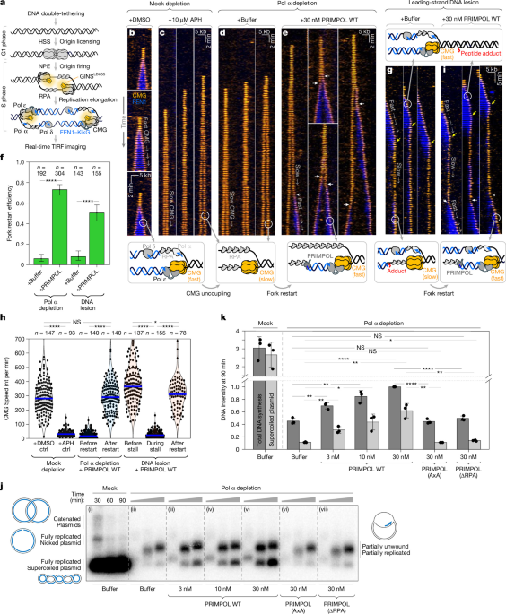
Eukaryotic genomes are exposed to numerous sources of DNA damage that impede replication. Only a decade ago, repriming by the primase–polymerase PRIMPOL was identified as a pathway that enables cells to tolerate DNA damage1,2,3,4. However, the molecular mechanism that underlies repriming remains poorly understood, and it is unclear how PRIMPOL is precisely targeted to stalled forks, where along single-stranded DNA PRIMPOL binds and how PRIMPOL is regulated to minimize error-prone DNA synthesis. Here, to dissect the molecular basis of repriming, we perform real-time single-molecule imaging in nuclear extracts that recapitulate genome maintenance. We directly visualize how stalled forks restart through repriming. We show that PRIMPOL is recruited to stalled forks through an interaction with the replicative helicase CDC45–MCM2–7–GINS (CMG). This facilitates primer handover from PRIMPOL to the high-fidelity polymerase ε, minimizing mutagenic DNA synthesis. At near-physiological concentrations, PRIMPOL reprimes leading strands in a CMG-dependent manner and polymerase ε extends those primers. This primary mode of repriming does not require DNA damage checkpoint signalling. PRIMPOL overexpression and checkpoint activation promote a secondary mode of repriming, whereby PRIMPOL restarts DNA synthesis far from CMG and polymerase δ extends those primers. Our data reveal a simple yet effective mechanism that directs PRIMPOL’s activity to stalled forks awaiting restart, and suggest a strategy to inhibit the repriming pathway.
Multiple pathways maintain genome stability during replication stress (RS)5, including fork reversal and translesion DNA synthesis (TLS)6. In 2013, a new RS response pathway called repriming was identified in eukaryotes1,2,3,4. PRIMPOL performs repriming and enables cells to tolerate diverse DNA lesions1,2,3,4,7,8. As it lacks proofreading, PRIMPOL has an error rate comparable to that of TLS polymerases (10−2−10−4)9,10. PRIMPOL-deficient cells and mice are hypersensitive to several DNA-damaging agents8,11,12,13, underscoring its role in genome maintenance. While repriming enables cancer cells to tolerate RS induced by oncogenes14 and chemotherapy15, PRIMPOL generates single-stranded DNA (ssDNA) gaps16,17 that sensitize cells to PARP inhibitors18,19,20. Blocking fork reversal enhances repriming, suggesting that cells favour high-fidelity pathways to minimize mutagenic DNA synthesis7,21,22.
The replicative helicase CMG unwinds parental DNA23 and is functionally coupled to polymerase ε (Pol ε)24, which synthesizes the leading strand. The lagging strand is primed and extended by primase–polymerase α (Pol α) and polymerase δ (Pol δ), respectively24. Damage on the lagging strand is readily bypassed due to the discontinuous nature of Okazaki fragment synthesis25,26. This is not the case for leading strand DNA lesions, which stall Pol ε while CMG continues to unwind DNA25,26,27. This CMG uncoupling exposes ssDNA that is rapidly coated by replication protein A (RPA)28. The notion that PRIMPOL may reprime leading strand synthesis was hypothesized at the time of its discovery2, and discussed in later work7,16,29, but there is no direct evidence for this idea.
Although repriming is a major RS response pathway and PRIMPOL is an attractive therapeutic target, key mechanistic aspects of repriming remain unclear. For example, although RPA recruits PRIMPOL to ssDNA30,31, it is unclear why PRIMPOL acts at stalled forks, but not on ssDNA generated by other processes (transcription, recombination). One study reported that high RPA concentrations inhibit PRIMPOL’s activity on ssDNA in vitro30, presumably because RPA fully coats ssDNA and prevents PRIMPOL’s access to the template. Another study found that RPA stimulated PRIMPOL’s activity on ssDNA long enough to accommodate the simultaneous binding of both proteins, that Pol ε can extend PRIMPOL’s primers in vitro and that RP