// NATURE NEWS — SPAZIO & SCIENZA
3D epigenome of glial cell types in developing human cortex
Nature
(2026) Cite this article
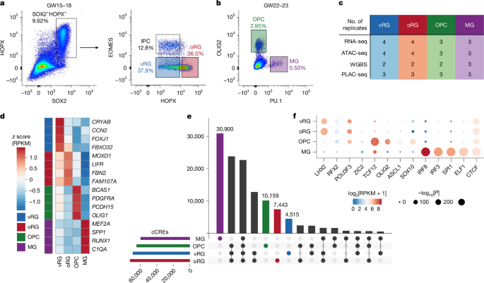
The human cortex is complex and heterogeneous, undergoing extensive expansion during development1,2. Our prior study of neurogenesis, including radial glia (RG), intermediate progenitor cells, excitatory neurons and interneurons demonstrated that chromatin looping underlies transcriptional regulation for lineage-specific genes, shedding light on how non-coding genetic variants contribute to neuropsychiatric disorders by means of cell-type-specific gene regulation3. RG have a crucial role in generating cellular diversity through both neurogenesis and gliogenesis and can be further classified into ventricular RG (vRG) and outer RG (oRG)4,5. Given their significance in cortical development, we conducted a comprehensive three-dimensional (3D) epigenomic analysis of four main glial populations, including vRG, oRG, oligodendrocyte precursor cells and microglia, from the mid-gestational human neocortex. By integrating gene expression, chromatin accessibility, DNA methylation and 3D chromatin interactions, we identified cell-type-specific candidate cis-regulatory elements (cCREs) and validated their regulatory function using transgenic mouse embryos. Using machine learning, we prioritized 112 schizophrenia risk variants within glia cCREs and further confirmed the predicted vRG enhancer disruption by the rs4449074 risk allele in vivo. Finally, oRG cCREs are enriched for human accelerated regions compared with other cCREs and a subset of human accelerated regions show activity differences from their chimpanzee orthologues that interact with genes involved in neuronal development. Our findings advance the understanding of human-specific gene regulation during corticogenesis.
The expansion of the human cortex is an extremely complex process that separates us from other mammals6,7. Radial glia (RG), the stem cells of the developing brain, are the driving force behind cortical expansion as they form a scaffold extending from the ventricular surface to the outermost pia that supports neuronal migration8. However, during mid-gestation, this scaffold becomes discontinuous as outer RG (oRG) delaminate from the ventricular surface, where ventricular RG (vRG) remain, and migrate towards the outer subventricular zone. In addition to different locations, vRG and oRG differ in the cell fate of their progeny5 and cell signalling pathways7,9. However, additional epigenomic profiling can provide further insight into mechanisms contributing to RG lineages and human-specific development, as much of our current knowledge is driven by transcriptional analysis. Although single-cell profiling of the developing brain highlights dynamic alterations of cell-type-specific epigenomes and transcriptomes10, the lack of integration of the three-dimensional (3D) epigenome with single-cell approaches poses substantial challenges to identifying mechanisms of epigenetic regulation, for example by chromatin loops, at the resolution required to analyse gene regulatory programs11,12. In addition, distal interacting regions detected by single-cell high-throughput chromatin conformation capture (Hi-C) tend to be less enriched for functionally validated enhancers compared with other modalities13, suggesting that these loops are more likely to represent structural interactions rather than promoter–enhancer loops.
Genome-wide association studies (GWAS) have identified thousands of variants associated with psychiatric disorders residing in non-coding regions, including cis-regulatory elements (CREs)14,15. Identifying causal variants remains challenging due to the heterogeneity of CREs across cell types9 as well as the difficulty of linking variants to their target genes as regulatory effects can span long genomic distances and do not necessarily affect the nearest gene16,17. Previously, we characterized cell populations involved in neurogenesis, including RG, intermediate progenitor cells (IPCs), excitatory neurons