// NATURE NEWS — SPAZIO & SCIENZA
A preinvasive regulatory T cell axis for lung cancer interception
Nature
(2026) Cite this article
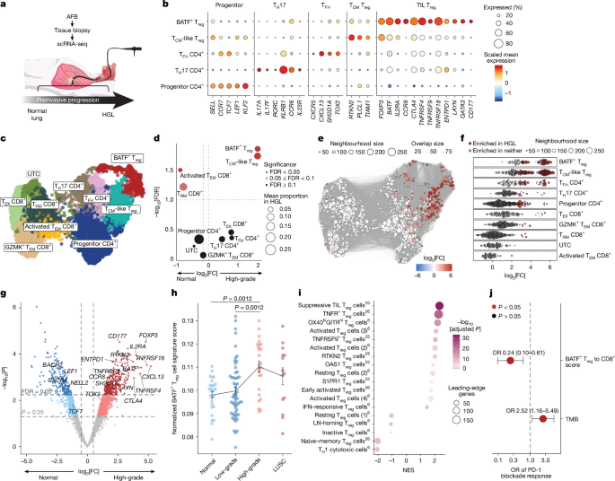
Late-stage non-small cell lung cancer (NSCLC) is rarely curable1, underscoring a need to intervene earlier in the disease process. Growing evidence suggests that antitumour T cell responses are mounted but become progressively dysregulated during early tumorigenesis2,3. Tracking and targeting preinvasive T cell regulation may inform new approaches to detect and intercept lung cancer development. Here we explore how the T cell network is remodelled during NSCLC development via multi-omic, cross-tissue immune profiling in patients with preinvasive lung lesions surveilled by autofluorescence bronchoscopy and computed tomography (CT) imaging. Effector regulatory CD4+ T cells (eTreg cells) expressing basic leucine zipper ATF-like transcription factor (BATF) accumulated in high-grade premalignant airway lesions and were clonally related to circulating eTreg cells. Circulating eTreg cells were increasingly elevated during preinvasive progression, enabling lung tumorigenesis to be tracked through analysis of peripheral blood. Emergence of this clonally coordinated eTreg cell circuit defined rapid progression in patients with early-stage NSCLC detected during CT screening. In mice, carcinogen-driven lung tumorigenesis triggered an analogous preinvasive eTreg cell axis across the blood, airways and draining lymph nodes (dLNs). This culminated in an expansion of lung BATF+ Treg cells and Treg cell-rich peribronchial immature tertiary lymphoid structures (iTLSs). Immune interception via phosphoinositide 3-kinase-δ (PI3Kδ) inhibition abrogated formation of Treg cell-rich iTLSs, reduced circulating and pulmonary Treg cells, increased local conventional type 1 dendritic cells (cDC1s) and reduced lung tumour incidence and size. These data reveal a conserved eTreg cell network that emerges across tissues during early pulmonary tumorigenesis and provide a theranostic framework to track and target preinvasive immune regulation for lung cancer interception.
NSCLC claims an estimated 1.8 million lives a year1. Despite substantial advances in early detection and therapy, prognosis remains poor, emphasizing the need to develop new prevention strategies. Lung cancer screening programmes are identifying an increasing number of preinvasive pulmonary neoplasia, which is creating an opportunity for NSCLC interception. However, clinical interception protocols remain underdeveloped owing to a limited knowledge of the biological pathways that drive tumorigenesis.
eTreg cells that express BATF (BATF+ Treg cells) define adverse survival and failure of neoadjuvant anti-PD-1 therapy in NSCLC4,5,6. Treg cells in NSCLC tumours share clonotypes with those in the circulation7, and their presence in tertiary lymphoid structures8 and tumour-draining lymph nodes (tdLNs)9 constrains antitumour immunity. Together, these results suggest that Treg cells form an immunoregulatory network that spans the circulation, lymphoid tissue and tumour in invasive NSCLC.
Growing evidence suggests that immune dysregulation precedes NSCLC invasion. Mouse models of lung adenocarcinoma (LUAD) development feature protumorigenic macrophage niches10, priming defects and CD8+ T cell dysfunction during early tumorigenesis11,12. Notably, preinvasive progression of human airway carcinoma in situ (CIS) lesions to lung squamous carcinoma (LUSC) is associated with impaired CD8+ T cell infiltration3, skewed CD4+ T cell differentiation and gene expression profiles emblematic of conventional T cell (Tconv cell) and/or Treg cell activation (for example, TNFRSF9 (encoding 4-1BB), ICOS, CTLA4, TNFRSF18 (encoding GITR), TGFB1 and IL10)13.
Here, we systematically explore T cell dynamics during clinical and experimental preinvasive pulmonary progression to identify immune regulatory pathways that could inform NSCLC interception. We discover a clonally coordinated cross-tissue eTreg cell circuit that progressively intensifies during carcinogen