// NATURE NEWS — SPAZIO & SCIENZA
Glycosylation and importins liquefy the FG barrier in live nuclear pores
Nature
(2026) Cite this article
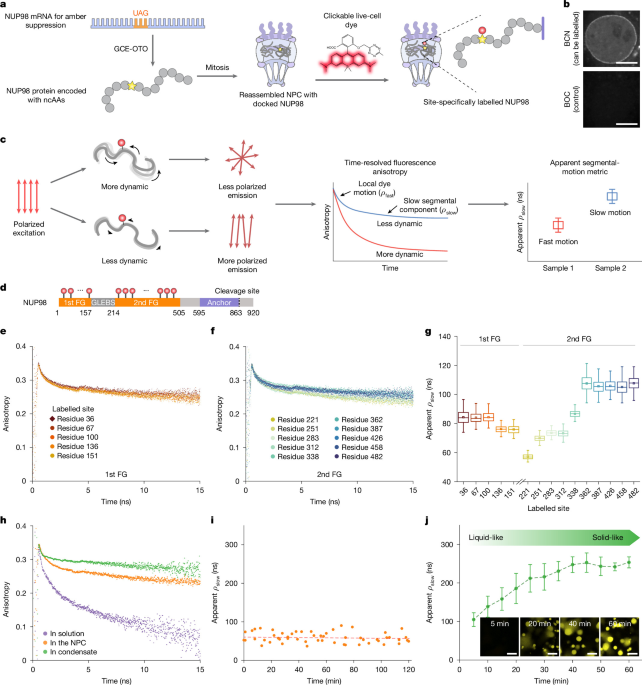
The nuclear pore complex (NPC) regulates molecular traffic between nucleus and cytoplasm1,2. Its permeability barrier is formed by intrinsically disordered phenylalanine-glycine-rich nucleoporins (FG-NUPs), whose physical state has long been debated3,4,5,6,7,8,9,10,11,12,13. Deciphering how FG-NUPs behave inside living cells is crucial for understanding how NPCs achieve both selective and rapid transport. By combining site-specific labelling with time-resolved fluorescence anisotropy, here we show that FG domains exhibit nanosecond-scale, liquid-like segmental motion in live cells, whereas reconstituted FG condensates undergo a liquid-to-solid transition in vitro. Experiments and molecular-dynamics simulations further show that importin-β, a principal nuclear transport receptor, and O-linked β-N-acetylglucosamine, a key post-translational modification of FG-NUPs, together stabilize the dynamic FG network. Finely balanced FG–FG and FG–importin-β interactions maintain FG-NUP liquidity and generate a radial organization in which importin-β is enriched near the NPC periphery and extended FG domains remain in the central channel, forming the transport barrier. This organization explains recent MINFLUX observations14 of rim-biased transport trajectories and is supported here by direct MINFLUX imaging of importin-β in mammalian cells. Together, our results reconcile conflicting models of FG-NUP organization4,5,7,9,15 and establish a framework for understanding how cellular context tunes intrinsically disordered proteins in health and disease.
The NPC mediates molecular exchange between the nucleus and cytoplasm. Its central permeability barrier comprises intrinsically disordered proteins (IDPs) known as FG-NUPs, which allow passive diffusion of small molecules while enabling the selective translocation of larger cargos bound to nuclear transport receptors (NTRs)1,2,16,17,18. How this functional barrier forms inside the nanochannel of the NPC remains unresolved.
Direct measurements of local FG-NUP conformational dynamics inside living NPCs remain challenging. Single-molecule tracking, fluorescence correlation spectroscopy and fluorescence recovery after photobleaching can probe overall diffusion in live cells19,20 but do not resolve local conformational motions in tethered disordered chains. Atomic force microscopy (AFM) has shown dynamic properties of NPCs and FG assemblies at accessible surfaces3,21 but is not readily applicable to intact NPCs in live cells. Consequently, models have relied heavily on in vitro experiments and simulations4,22,23, ranging from flexible polymer chains24,25 and polymer brushes15 to liquid-, gel- and amyloid-like condensates4,5,11,12,13. Whether the native barrier is predominantly liquid-like or more solid-like therefore remains debated.
Our previous fluorescence lifetime imaging microscopy–Förster resonance energy transfer (FLIM–FRET) measurements defined conformational preferences of FG-NUPs in permeabilized cells26 but did not directly report dynamics in live NPCs. We therefore combined site-specific genetic-code-expansion labelling with picosecond time-resolved fluorescence anisotropy (TRFA) to monitor apparent segmental motion in live mammalian cells. Focusing on the vital FG-NUP98, we find that FG domains remain liquid-like in living cells, whereas reconstituted FG condensates undergo a time-dependent liquid-to-solid transition in vitro.
We further identify importin-β, an abundant NTR, and O-linked β-N-acetylglucosamine (O-GlcNAc), a key post-translational modification (PTM) of metazoan FG-NUPs, as cellular factors that tune FG-domain liquidity. In vitro, either factor suppresses FG-NUP ageing, whereas together they closely reproduce the apparent segmental motion observed in cells. In live cells, reduced cellular O-GlcNAcylation slows apparent FG-domain segmental motion and nuclear transport, whereas importin-β overexpression partial