// NATURE NEWS — SPAZIO & SCIENZA
Luminescent-reaction-enabled super-resolution imaging
Nature
(2026) Cite this article
By breaking the optical diffraction limit, super-resolution fluorescence microscopy has advanced our understanding of biological complexity under the framework of light-excited luminescence1. The use of external light excitation remains a key factor that shapes the imaging capabilities and live-cell compatibility of fluorescence-based approaches2. An alternative is the reaction-excited luminescence, such as electrochemiluminescence (ECL)3, chemiluminescence (CL)4 and bioluminescence (BL)5, providing a chemically defined toolbox for enabling different imaging merits, from ultrasensitive analysis6,7 to biocompatible imaging8,9. Despite its light-free excitation and high sensitivity, conventional luminescent-reaction-enabled imaging is fundamentally limited in spatiotemporal resolution owing to low photon budget10,11. Here we develop a chemistry-based super-resolution imaging framework, luminescent-reaction-enabled super-resolution imaging via entropy-weighted correlation combined with deconvolution (RIED). As an experimental–computational concept, RIED introduces a spatiotemporal recording strategy to uncover specific luminescent-reaction-enabled imaging information content, which is efficiently collected and computed to achieve super resolution using a reconstruction strategy adapted to reaction-driven photon statistics. We achieve super-resolution ECL, CL and BL imaging of intracellular organelles, attaining approximately 100 nm resolution. This approach is used for highly sensitive imaging of surface proteins and 41-h ultralong-term continuous super-resolution live-cell imaging of mitochondrial transfer dynamics. Our work establishes an emerging class of chemistry-enabled, laser-free super-resolution microscopy with expanded biological imaging versatilities.
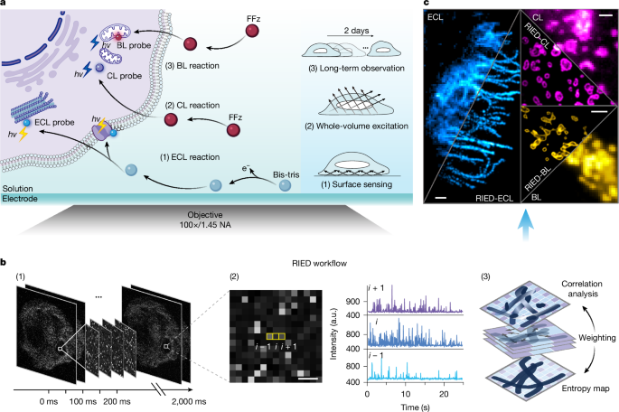
Reaction-enabled imaging is based on specifically designed luminescence chemistry, which can be tailored for different applications (Fig. 1a). Specifically, ECL is triggered and controlled by electrochemical reactions, imparting high surface sensitivity, which inherently facilitates imaging of objects near the electrode. CL relies on the reactant diffusion in solution, enabling homogeneous excitation throughout the imaging volume. BL makes use of enzymatic reactions to produce catalytic luminescence in living organisms, providing excellent biocompatibility for live-cell imaging. Despite these advantages, so far, the reaction-based luminescence suffers from low photon output, usually requiring prolonged exposure times by tens of seconds to accumulate a single image10,11, which inevitably overlooks the spatiotemporal information. As a result, unlike established super-resolution fluorescence microscopy, achieving super resolution using reaction-enabled luminescence remains challenging.
a, Schematic of luminescent-reaction-based imaging—ECL, CL, BL and their features. b, Workflow of RIED reconstruction: (1) spatiotemporal acquisition of luminescent signals; (2) zoomed photon-signal distribution for a single frame in (1) and corresponding intensity profiles of three separate pixels highlighted by yellow boxes; (3) schematic of the core reconstruction principle used in RIED. Spatiotemporal correlation and entropy maps are calculated and weighted for super resolution. c, Representative super-resolution ECL, CL and BL images of intracellular organelles before and after RIED reconstruction. Scale bars, 3 µm. a.u., arbitrary units.
From a fundamental perspective, the reaction-based luminescence is a highly dynamic process, intrinsically initiated by individual reactions. To achieve super-resolution imaging, we propose to experimentally monitor these reaction-based luminescence processes in an image sequence through a spatiotemporal isolation strategy. This enables the extraction of distinct reaction-driven photon statistics for super-resolution reconstruction using a tailored workflow (Fig. 1b, Extended Data Fig. 1 an