// NATURE NEWS — SPAZIO & SCIENZA
Functional role of skull lymphoid structures in CNS immunosurveillance
Nature
(2026) Cite this article
Accumulating evidence demonstrates that the central nervous system (CNS) is not disconnected from the peripheral immune system; however, precisely how the adaptive immune system surveils the CNS remains a critical question. Recent findings reveal that channels between the skull and the dura mater facilitate the exchange of cerebrospinal fluid and immune cells between the CNS and skull bone marrow of mice under both homeostatic and disease conditions1,2,3,4,5,6. Skull bone marrow functions as a source of immune cells for the CNS5, yet its role in CNS antigen-specific adaptive immune responses remains unclear. Here we identify lymphoid structures within the skull bone marrow, featuring germinal-centre-like formations and containing a distinct population of follicular-helper-like T cells that promote B cell activation and humoral immunity through CD40L, IL-21 and IFNγ signalling. Adaptive immune cells within these skull bone marrow lymphoid structures surveil and respond to CNS-derived antigens and contribute to anti-tumour immune responses in mouse brain cancer models. Together, our findings show that the skull bone marrow is a site of CNS immunosurveillance that may influence immune responses across diverse neurological diseases.
Recent findings have highlighted the direct connection between skull bone marrow (BM) and the dura mater through osseous channels, enabling cerebrospinal fluid (CSF) to reach the skull BM1,2,3,4,7. This connection is functionally important, as the skull BM is a primary lymphoid organ that supplies immune cells—including developing and immature B cells and myeloid cells—to the CNS and CNS border regions under homeostatic and neuroinflammatory or injurious conditions5,6. Recent studies also suggested that skull BM contains tumour-specific CD8+ T cells in human patients with glioblastoma and regulatory T cells in mouse tumour models8,9. Although the BM is a primary lymphoid organ, adaptive immune responses also occur within it under certain conditions10,11,12. Mature antigen-presenting cells (APCs), T cells and B cells migrate to the BM, where residing naive T and B cells can encounter antigens and form clusters13,14,15,16,17,18,19. While previous studies have shown that BM can support adaptive immune responses, whether and how skull BM contributes to CNS antigen recognition and immunosurveillance has not yet been elucidated.
Here we demonstrate that the skull BM of mice contains cellular components that are characteristic of peripheral lymphoid organs. APCs, T cells and B cells form clusters with germinal centre (GC)-like structures. These skull lymphoid structures participate in CNS immune surveillance under both homeostatic and brain cancer conditions in mice. By modulating their presence and activity in vivo, we show that these structures contribute to anti-tumour immunity.
We compared T cell phenotypes in the skull BM to those of other BM sites in mice to characterize the adaptive immune composition of skull BM. The frequency of CD4+ and CD8+ T cells within total immune cells was similar across BM sites, with a higher frequency in the sternum BM, but overall lower than in secondary lymphoid organs (SLOs) (Extended Data Fig. 1a,b). The frequencies of naive and effector T cells were also comparable. However, skull BM displayed a higher proportion of central memory cells compared with other BM sites (Extended Data Fig. 1c,d), suggesting a distinct T cell environment within the skull BM.
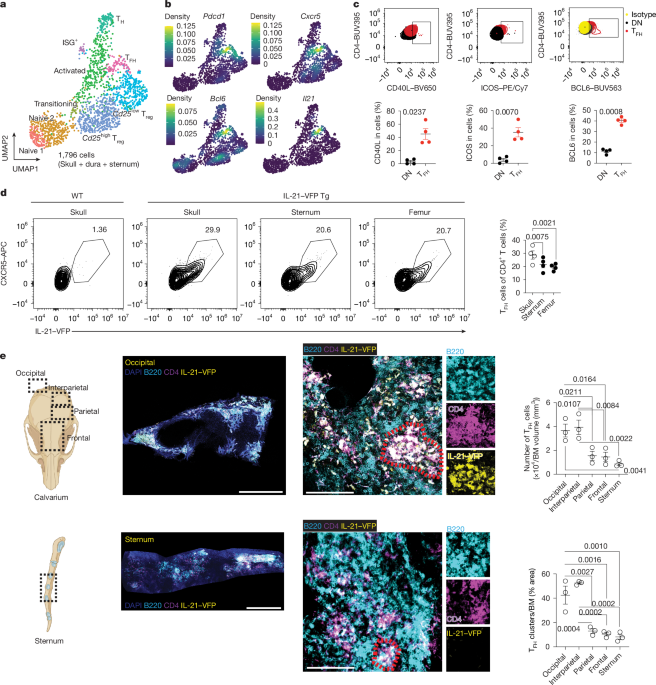
To further examine T cell phenotypes in BM, we conducted 10x single-cell RNA sequencing (scRNA-seq) analysis of T cells from mouse skull and sternum BMs. On the basis of clustering, we identified diverse CD4+ T cell subsets, including naive (Sell and Ccr7), activated (Cd44), interferon-stimulated gene (ISG)-expressing (Isg15), regulatory (Foxp3) and differentiated helper (Tbx21, Gata3 and Rorc) cells (Fig. 1a and Extended Data Fig. 1e). Notably, we observe