// NATURE NEWS — SPAZIO & SCIENZA
Proximity-guided graph learning reveals tumour-associated proximity antigens
Nature
(2026) Cite this article
The spatial organization of membrane proteins is an underexplored dimension of cell surface biology1,2. Spatial proximity shapes cellular function and therapeutic targetability2,3, yet efforts to identify tumour-associated antigens (TAAs) have largely focused on expression alone4. Here, we developed an industrialized surface protein proximity-mapping workflow to interrogate TAAs within their membrane microenvironments. Using this workflow, we generated 248 proximity maps across 12 receptor tyrosine kinases and 28 tumour cell systems. The resulting atlas enabled the development of MetaMap, a correlation-based analytical framework that defines spatial protein communities and infers conserved proximity relationships among non-targeted proteins, and establishes the concept of tumour-associated proximity antigens (TAPAs), a class of co-targets defined by disease-specific spatial proximity to TAAs rather than expression alone. Integrating these proximity-derived relationships within a multimodal prioritization framework, we identified and validated EGFR–CDCP1 as a TAA–TAPA pair that enhances tumour cell killing across therapeutic modalities. Together, this work advances disease-associated membrane proximity as a guiding principle for the design of precision multispecific therapeutics.
The cell surface is a dynamic landscape where membrane protein organization regulates diverse biological processes, including immune synapse formation, epithelial junctional organization and receptor-mediated signalling1,5. Membrane protein proximity is a defining feature of immune receptors6, cytokine receptors7 and receptor tyrosine kinases (RTKs)3, many of which assemble into localized signalling hubs2.
In cancer, the cell surface proteome is remodelled through changes in protein abundance, trafficking, post-translational modifications, mutations and membrane organization4,8,9,10. These changes reshape surface protein spatial networks, disrupting normal physiology and promoting disease progression8,9,10,11,12. Notably, this protein-level spatial organization is not captured by gene or protein expression analyses, which measure abundance rather than proximity. Resolving membrane protein organization is therefore critical for contextualizing TAAs within functional neighbourhoods and guiding multispecific therapeutic design.
Despite the importance of spatial organization, scalable approaches for mapping surface protein microenvironments remain limited. We previously introduced complementary photocatalytic proximity-labelling technologies that covalently label proteins within a nanoscale radius to map membrane microenvironments13,14,15,16. In this Article, we integrate these orthogonal chemistries into an industrialized high-throughput microenvironment mapping (micromapping) workflow. Because labelling is governed by reactive intermediates with finite lifetimes and diffusion distances, these measurements capture local membrane neighbourhoods rather than only direct physical interactions. The workflow incorporates standardized procedures and cross-experiment normalization to quantitatively compare protein proximity across targets, cell systems and tumour contexts.
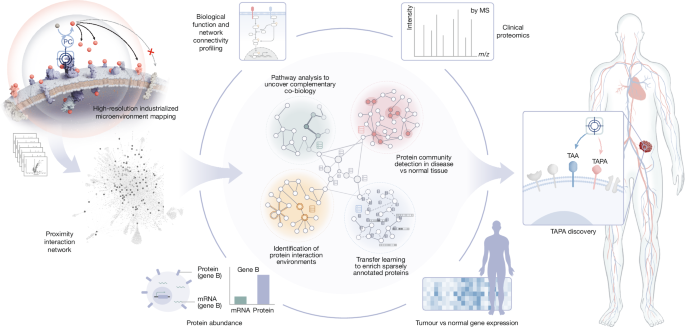
Using this platform, we mapped surface protein microenvironments across diverse cancer models. To extend these measurements beyond directly targeted proteins, we developed MetaMap, an analytical framework that defines spatial protein communities and infers conserved proximity relationships among non-targeted proteins. We then applied graph-based learning informed by protein proximity and abundance to identify candidate co-target pairs, which were subsequently prioritized using normal tissue expression, tumour-versus-normal expression, clinical proteomics and disease-relevant signalling features (Fig. 1). This approach prioritizes proximity-defined co-targets by revealing which TAAs are organized together on tumour cells, rather t