// ARS TECHNICA — MONDO
Finding the cells that put our brain to sleep
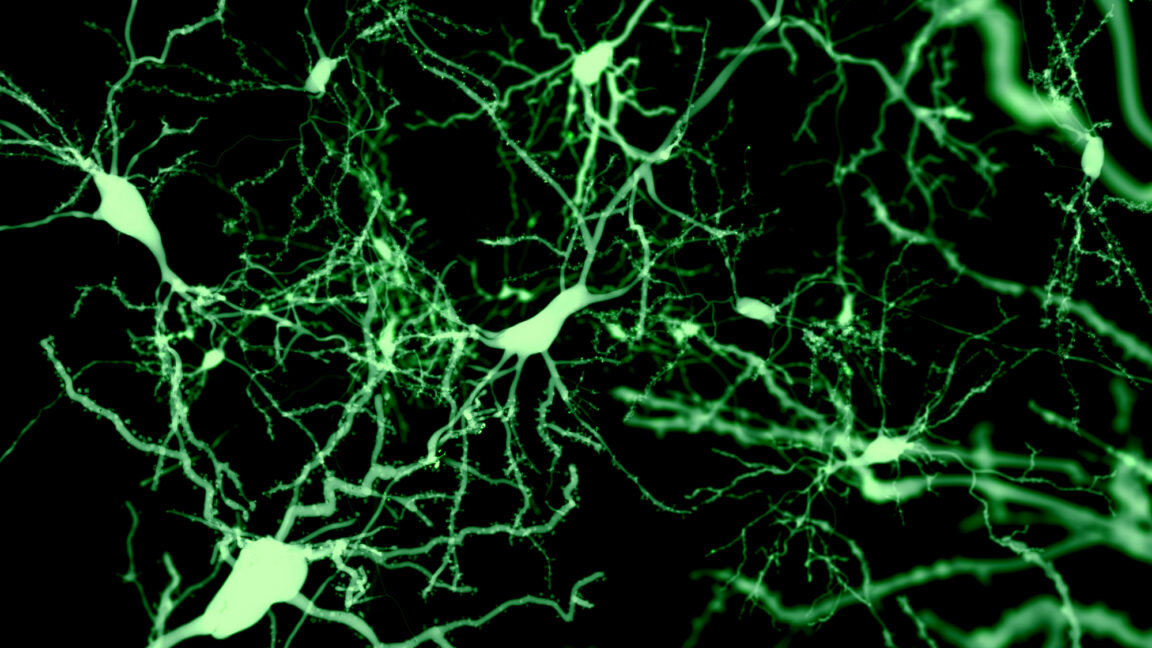
Rare cells with widespread connectivity seem to be able to trigger sleep.
For a long time, sleep research has treated the cerebral cortex as a passive follower reacting to signals from the deep brain. “Usually, sleep is associated with being controlled by subcortical regions,” said Geoffrey Terral, a neuroscientist at the Albert Einstein College of Medicine in New York.
The cortex is where the slow rhythms of deep sleep can be seen, but researchers assumed the signals that triggered them originated elsewhere. In a recent Nature study, Terral and Renata Batista-Brito, who runs the lab, report a population of cortical cells that challenges that assumption.
These cortical cells make up only around one percent of the cortex’s inhibitory neurons, and switching them on in a mouse puts the animal to sleep. “What our work shows is that the cortex can not only see this rhythm but also initiate it by itself, and this is sufficient to promote sleep,” Terral said.
The cells are called Sst-Chodl neurons, after two genes that are active in them. Long-range inhibitory neurons marked by the activity of these genes have been known for some time, based on studies in monkeys, but researchers couldn’t figure out how to manipulate them using these genes. Aiming at either one of these genes captures a huge, heterogeneous family of different neurons. “A single gene is not able to target these cells,” Batista-Brito said.
Inhibitory neurons account for about a fifth of all cortical neurons, so Sst-Chodl cells amount to roughly one neuron in a thousand in the cortex. “If you’re not really specific, the contaminants are going to be much more dominant than the specific cells,” Batista-Brito said.
Building a strategy that labels a cell only when both genes are on took her team years.
“Going after these cells was really a high-risk project because the likelihood of seeing anything with 0.1 percent of neurons in the cortex is really low,” Batista-Brito said. “I wrote a bunch of grants on these projects that were always rejected because it was too high-risk.”
Her first look at the anatomy of Sst-Chodl neurons proved the risk worth taking. “At first, I saw two or three cell bodies in the whole brain,” Batista-Brito told Ars. “Despite that, there was massive, massive arborization all over the visual cortex, like I never saw with any other neuron.”
That “massive arborization” is a tree-like branching that connects neurons to other parts of the brain, making them part of a wider communication network. It was also very unusual, as inhibitory neurons are almost always local. They receive inputs from different, sometimes distant regions in the brain and exert control over their immediate surroundings—their own small patch.