// NATURE NEWS — SPAZIO & SCIENZA
Hepatocytes promote liver metastasis of pancreatic cancer by providing serine
Nature
(2026) Cite this article
The liver is the primary site of metastasis in pancreatic ductal adenocarcinoma (PDAC), and liver metastases are a major cause of mortality1,2. Nutrient availability in the metastatic niche influences colonization efficiency; however, the metabolic heterogeneity of disseminated tumour cells can also reshape the local microenvironment3,4,5. Loss of phosphoglycerate dehydrogenase (PHGDH), the rate-limiting enzyme in de novo serine biosynthesis, is observed in nearly 40% of PDACs, and renders these cells dependent on exogenous serine (exSer)6. Although a neuron–tumour metabolic cross-talk supports exSer-dependent PDAC cells at the primary site6, it remains unclear how these cells adapt to the metastatic liver niche. Here we show that exSer-dependent PDAC cells reprogram neighbouring hepatocytes through a CXCL5–CXCR2 axis. Activation of CXCR2 in hepatocytes promotes PI3K–AKT signalling, leading to the sequestration of FOXO3A in the cytoplasm and derepression of PHGDH transcription, thereby enhancing serine production in hepatocytes. This hepatocyte-derived serine supports the outgrowth of exSer-dependent PDAC liver metastases. Accordingly, genetic or pharmacological inhibition of individual nodes within the CXCL5–CXCR2–PI3K–AKT–FOXO3A axis, or hepatocyte-specific deletion of Phgdh or Cxcr2, markedly reduces the liver-metastasis burden in mice and prolongs survival, particularly when dietary serine is restricted. Our findings reveal a cancer cell–hepatocyte metabolic cross-talk and identify therapeutic targets for exSer-dependent PDAC liver metastases.
Previously, we reported that 37.2% of primary PDAC tumours exhibit a loss of PHGDH expression6. These PHGDH-deficient PDAC cells lack the capacity for de novo serine synthesis and therefore rely on exogenous serine (exSer) for their survival and proliferation. In the primary tumour setting, these exSer-dependent PDAC cells use serine that is released from sensory nerves, and this constitutes a distinct and therapeutically targetable metabolic vulnerability6. However, it remains unclear how exSer-dependent PDAC cells obtain serine in the metastatic setting, and particularly in liver metastases, where the tumour microenvironment (TME) differs markedly from that of the primary tumour7. This lack of knowledge is due in part to a limited availability of clinical specimens, because patients with liver metastases are ineligible for surgical resection1.
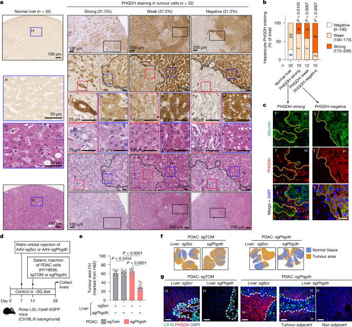
To examine the serine synthesis pathway (SSP) in PDAC liver metastases, both in cancer cells and in the surrounding metastatic microenvironment, we analysed the expression of PHGDH in human PDAC liver-metastasis samples collected through a rapid-autopsy programme8. We found that 10 out of 32 cases (31.3%) were PHGDH-negative in tumour cells (Fig. 1a,b), a frequency comparable to that observed in primary tumours6. Notably, hepatocytes adjacent to metastatic lesions, particularly those with weak or negative expression of PHGDH in tumour cells, exhibited strong PHGDH expression, whereas hepatocytes in non-metastatic liver tissue showed weak or negative staining (Fig. 1a,b). Dual immunofluorescence staining for PHGDH and albumin (a hepatocyte marker) confirmed this reciprocal pattern, with strong PHGDH expression in hepatocytes adjacent to PHGDH-negative PDAC metastases but little or no PHGDH expression around PHGDH-strong metastases (Fig. 1c). To validate this finding in vivo, we established liver metastases in nude mice using human PDAC cell lines with differential PHGDH expression: MiaPaCa2 (PHGDH-high) and PaTu8902 (PHGDH-null)6. Consistent with the human data, dual immunofluorescence staining revealed minimal expression of PHGDH in hepatocytes surrounding MiaPaCa2 metastases but a marked upregulation of PHGDH in those adjacent to PaTu8902 metastases (Extended Data Fig. 1a). Given the central role of hepatocytes in maintaining metabolic homeostasis, we hypothesized that hepatocyt