// NATURE NEWS — SPAZIO & SCIENZA
Dynamics of human cardiogenesis and its disruption in trisomy 21
Nature
(2026) Cite this article
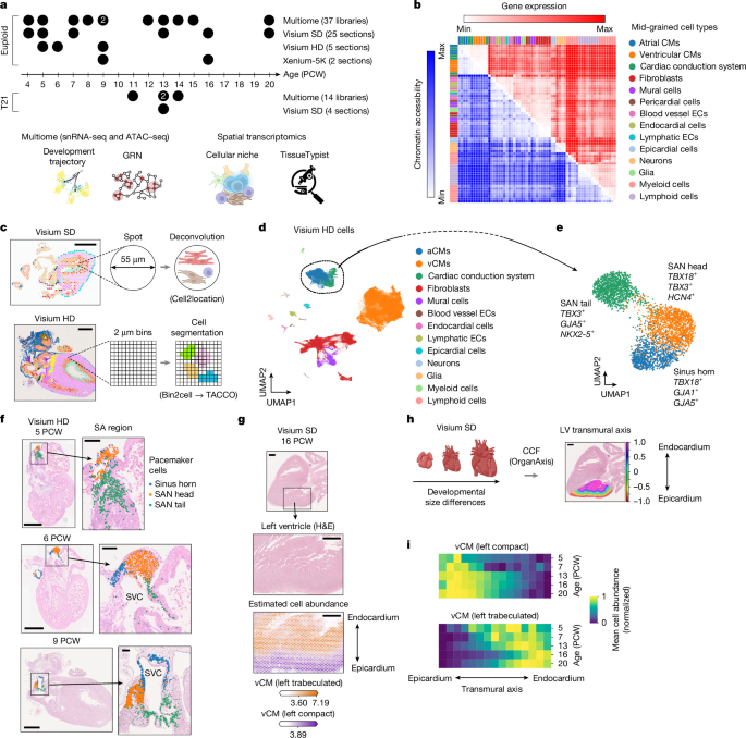
Developmental dynamics involve the specification of diverse cell types and their spatial organization into multicellular niches1. Here we combine single-cell and spatial multiomics to define 21 distinct tissue niches in the developing heart, which we use to develop a context-aware, resolution-agnostic niche classification tool (TissueTypist). Applying high-resolution spatial profiling to the developing sinoatrial node, we resolve three pacemaker cell subtypes arrayed along a linear axis. First trimester subpopulations, such as pacemaker cells in the sinus horn and sinoatrial node head region, display neuroattractant programs and interact with parasympathetic neurons via interactions that include Eph–ephrin and semaphorin–plexin signalling. Temporal trajectories map the maturation of atrial and ventricular cardiomyocytes and uncover a lipid–metabolic switch and potential key regulators of cell-type identity. In the ventricle, we identify cellular and transcriptional gradients along both pseudotime and transmural axes, which provide molecular insights into myocardial compaction and maturation. Comparative profiling revealed that hearts with trisomy 21 are depleted in compact cardiomyocytes and exhibit increased apoptosis relative to euploid hearts. This finding was validated in isogenic-matched trisomy 21 and euploid cardiomyocytes derived from induced pluripotent stem cells. These early developmental perturbations may contribute to the increased risk of congenital heart disease associated with Down’s syndrome. In summary, we present a spatially resolved framework of human cardiac development to enable systematic explorations of developmental niches in health and disease.
Human cardiogenesis requires coordinated interactions among diverse cell types that originate from the cardiogenic mesoderm and neural crest. These populations orchestrate key processes such as formation of the sinoatrial node (SAN), the primary pacemaker of the heart, and maturation of cardiomyocytes1,2. These mechanisms depend on precise spatial and temporal organization of cell states and their integration into multicellular niches that guide morphogenesis and functional maturation. Although genetic, epigenetic and environmental perturbations can disrupt these processes, which can lead to congenital heart disease (CHD), the underlying cellular and spatial mechanisms remain incompletely understood. Notably, the markedly increased incidence of CHD in trisomy 21 (T21) highlights the sensitivity of cardiac development to altered gene dosage3. Despite the identification of numerous CHD-associated genes, a comprehensive view of how early molecular and cellular perturbations emerge and are organized during human heart development remains lacking.
Although cardiomyocytes derived from human pluripotent stem cells have provided insights into aspects of cardiomyocyte identity and maturation4,5,6, they do not fully recapitulate in vivo development, particularly with respect to tissue organization and cellular diversity. Recent efforts to profile the fetal heart using transcriptomic and spatial omics approaches have revealed spatial gene expression patterns and cell-type heterogeneity7,8,9,10. However, these studies have generally been limited in developmental coverage, spatial resolution or modality of data types. Furthermore, robust methodologies for the identification and annotation of cellular niches in the developing heart, using various resolutions of spatial omics data, have yet to be established. Consequently, there remains a clear need for comprehensive, multimodal and spatially resolved atlases that can be used to analyse the cellular and molecular dynamics of human cardiac development.
Here to address these knowledge gaps, we construct a spatially resolved multimodal atlas of the developing human heart from 4 to 20 post-conceptional weeks (PCW), integrating single-cell transcriptomic data11 with