// NATURE NEWS — SPAZIO & SCIENZA
NRF2 is a mechanosensitive checkpoint for NLRP3 inflammasome activation
Nature
(2026) Cite this article
Disconnects between in vitro and in vivo results in experimental biology have long been recognized as potential hurdles to understanding health and disease. A prototypical example of such a disconnect is the NLRP3 inflammasome, which mediates inflammation in diverse settings of disease and protective immunity1,2. Activation of the NLRP3 inflammasome in vitro requires two signals: (1) priming by pathogen-associated molecular patterns such as bacterial lipopolysaccharides and (2) activation by molecules that disrupt cellular homeostasis1,3. By contrast, the NLRP3 inflammasome in vivo is activated by lipopolysaccharide alone. The mechanisms that underlie the difference between in vitro and in vivo activation of the NLRP3 inflammasome are undefined. Here we report that extracellular matrix (ECM) stiffness is a determinant of NLRP3 activities and that in vitro systems using physiological ECM stiffness enable one-signal in vivo-like NLRP3 activities to occur in human and mouse phagocytes. Physiological ECM stiffness induced expression of the transcription factor NRF2, which was found to be necessary for one-signal NLRP3 activation. Mechanistically, we identify Panx1 as an NRF2-regulated gene that promotes NLRP3 activity. Macrophages derived from the stiff matrix of the bone—osteoclasts—had reduced NRF2 levels and impaired NLRP3 activity. Moreover, alveolar macrophages from patients with pulmonary fibrosis displayed reduced NRF2 activity. Finally, NRF2 was required for antitumour immunity induced by cancer vaccines consisting of NLRP3 agonistic adjuvants. These findings indicate that NRF2 may be a transcriptional checkpoint that controls NLRP3 inflammasome activity.
Inflammasomes are cytoplasmic organelles that mediate inflammatory responses to infection and injury, primarily through the cleavage and release of the interleukin-1 (IL-1) family of cytokines3. Diverse proteins can seed inflammasome assembly, with NLRP3 being the best characterized and most commonly associated with human disease. NLRP3 activation in vitro requires two signals1. Signal 1 is provided by pathogen-associated molecular patterns such as bacterial lipopolysaccharide (LPS), which sensitizes (that is, primes) the NLRP3 pathway for responsiveness to subsequent stimuli. Signal 2 can be provided by a diverse set of stimuli, all of which induce interactions between NLRP3 and the protein ASC, which leads to the recruitment and activation of caspase-1 (refs. 1,4). The NLRP3–ASC–caspase-1 complex represents the minimal components of the NLRP3 inflammasome. Activated caspase-1 in the inflammasome cleaves pro-IL-1β and the pore-forming protein gasdermin D (GSDMD), which leads to the release of bioactive IL-1β (IL-1β p17) and pyroptosis5,6,7,8.
Although this two-signal model holds true for NLRP3 activation in vitro, LPS alone is sufficient to activate NLRP3 in vivo9,10,11,12,13,14,15,16. An explanation for the discrepancy between in vitro and in vivo NLRP3 activation remains elusive and was investigated herein.
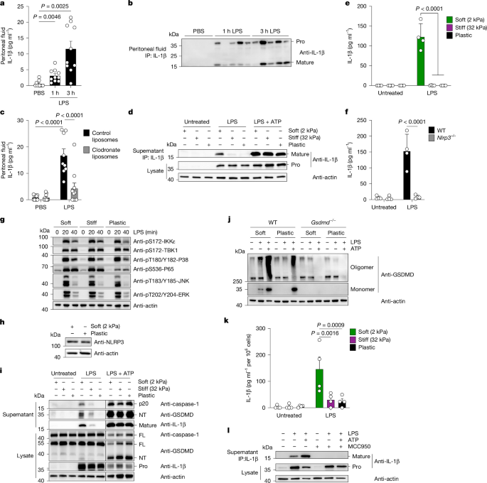
We investigated the kinetics of NLRP3 activation in vivo after intraperitoneal (i.p.) injection of LPS into mice. LPS injection induced the NLRP3-dependent accumulation of IL-1β in the serum within 3 h (Extended Data Fig. 1a,b). Caspase-11 is a regulator of LPS-induced IL-1β production in vivo. Caspase-11 was dispensable for IL-1β accumulation in the serum 3 h after LPS injection but was required for IL-1β production 6 h after LPS injection (Extended Data Fig. 1c). These data indicate that LPS can activate NLRP3 rapidly and independently of caspase-11. Within 1 h of LPS injection, cleaved IL-1β was detected in the peritoneal fluid (Fig. 1a,b). Nlrp3−/− mice did not accumulate IL-1β in the peritoneal fluid after LPS injection (Extended Data Fig. 1d). The speed of IL-1β p17 production observed in the peritoneal fluid suggested that a pool of resident cells is responsible. We depleted peritoneal macrophag