// NATURE NEWS — SPAZIO & SCIENZA
Intracellular complement factor H protects neurons during CNS inflammation
Nature
(2026) Cite this article
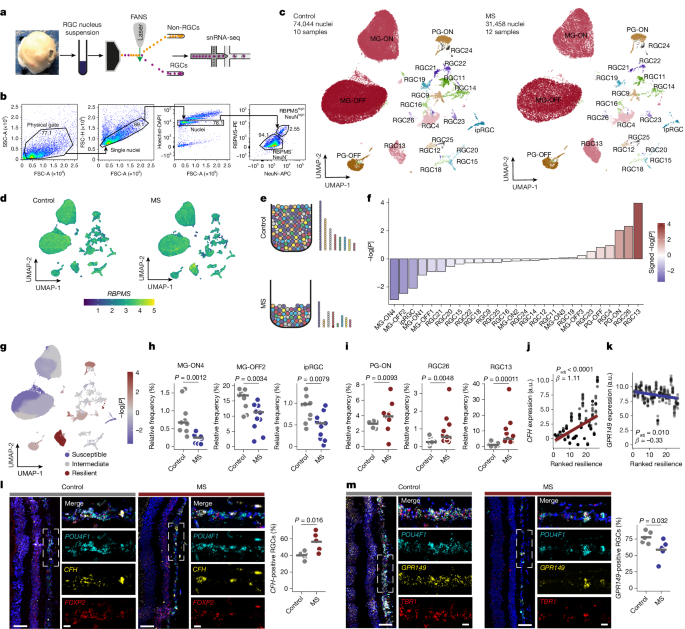
Neurodegeneration is a major driver of disability in multiple sclerosis (MS), the most common chronic inflammatory disease of the central nervous system (CNS)1. Retinal ganglion cells (RGCs), a heterogeneous neuronal population in the eye, undergo degeneration in MS and provide a model to study neuronal subtype-specific resilience to inflammatory injury2. However, the neuron-intrinsic mechanisms underlying differential vulnerability remain unclear. Here we identify a neuroprotective role for intracellular complement factor H (CFH) in neurons. Using single-nucleus RNA-sequencing analysis of RGCs from donors with MS and control individuals, we found that CFH expression was strongly correlated with intrinsic resilience to RGC degeneration. Mechanistically, CFH was induced in retinal and other CNS neurons in response to inflammatory and oxidative stress, where it limited reactive oxygen species accumulation and lipid peroxidation. CFH localized to the endoplasmic reticulum, a major site of lipid peroxidation during neuronal ferroptosis. Its protective activity was dependent on its C-terminal SCR20 domain, was independent of CFH secretion and was preserved in the absence of complement component C3. These findings reveal a non-canonical intracellular function of CFH in neurons. Together, our results identify CFH as a key mediator of neuronal resilience across the CNS in mice and humans and provide mechanistic insight into inflammatory neurodegeneration with implications for MS therapy and neuroprotection more broadly.
In MS, a disease that predominantly affects young adults, inflammation is followed by early and widespread neurodegeneration throughout the CNS1. The disease is thought to be driven by autoreactive T cells infiltrating the CNS and triggering chronic myeloid cell activation that sustains low-grade inflammation and gradually drives neurodegeneration across CNS regions, including the eye3,4. Although several immunomodulatory therapies target the peripheral immune system, none address the CNS-intrinsic inflammatory environment or reinforce stressed neurons against inflammation-induced degeneration, limiting their impact on progressive disability5.
In addition to motor and sensory impairments, visual disturbances are among the most common presenting symptoms in people with MS4. Retinal atrophy is an early feature of MS and can be detected even without a previous episode of optic neuritis6. Thus, acute and chronic low-grade CNS inflammation leads to substantial degeneration of RGCs, the output neurons of the retina of which the axons form the optic nerve.
RGCs are among the most diverse neuronal populations, with over 40 distinct subtypes described in mice2. This diversity has driven investigations into subtype-specific vulnerability, particularly in models such as optic nerve crush, where around 80% of RGCs degenerate within the first 2 weeks2. Notably, because humans evolved a fovea for high-resolution colour vision, RGCs in this region may face a trade-off of increased susceptibility to degeneration, as seen in age-related macular degeneration (AMD) and MS, emphasizing the need to study the mechanisms of retinal degeneration in humans7.
Recent work has begun to resolve the transcriptional heterogeneity of human RGCs8,9,10, but cell type identification remains inconsistent and datasets are dominated by midget RGCs, limiting analysis of rarer subtypes. Although MS substantially affects the retina, single-cell studies have focused largely on the brain11,12,13 and histopathological studies of the MS retina are sparse14, leaving a gap in our understanding of how MS-associated inflammation affects retinal neurons. Moreover, most single-cell studies describe molecular changes without translating them into clinical concepts or therapies. To address this, we performed single-nucleus sequencing analysis of RGCs from control and MS retinas to identify genes determinin