// NATURE NEWS — SPAZIO & SCIENZA
MMEJ repair of breaks at TA repeats maintains ecDNA and cancer fitness
Nature
(2026) Cite this article
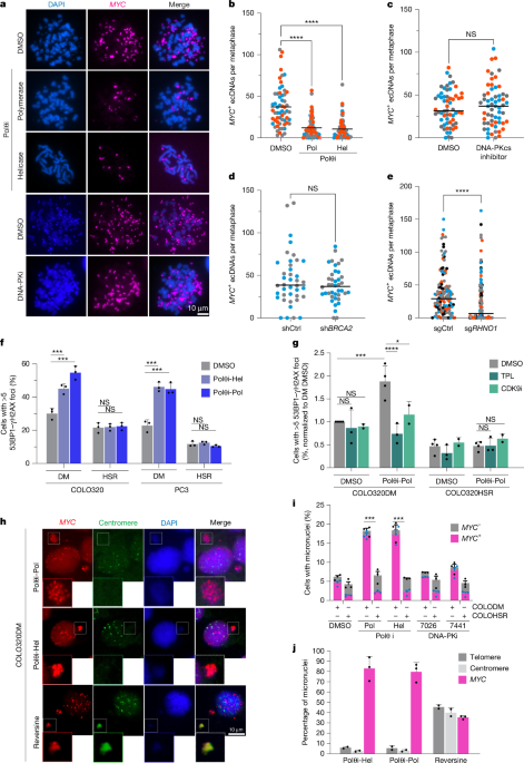
Extrachromosomal DNA (ecDNA) comprises megabase-sized circular DNA elements that frequently carry oncogene amplifications, driving aggressive tumour phenotypes, therapeutic resistance and poor clinical outcomes across many cancers1,2,3,4. Although ecDNA is thought to arise from canonical double-strand break repair, the pathways that maintain it remain unclear. Here we show that inhibition of microhomology-mediated end joining, but not non-homologous end joining or homologous recombination, selectively depletes ecDNA, induces ecDNA-specific damage and promotes its sequestration into micronuclei, compromising the fitness of cancer cells that depend on ecDNA-driven oncogene amplification. Mechanistically, TA-rich loci on ecDNA are hotspots for DNA damage and breakage5,6. The DNA translocase FANCM suppresses break formation at these sites, while breaks that escape FANCM surveillance are cleaved by ERCC1–ERCC4 and channelled into microhomology-mediated end joining for repair. Single-cell whole-genome sequencing shows that disrupting FANCM or polymerase θ (Polθ) in COLO320DM cells causes structural instability characterized by deletions and small duplications, with breakpoints enriched at TA-rich regions. This fragility is recapitulated in human tumours, in which ecDNA rearrangements are enriched at TA repeats. Collectively, our findings reveal TA repeat fragility as an intrinsic vulnerability of circular DNA and identify Polθ inhibition as a promising strategy to potentially destabilize ecDNA and sensitize ecDNA-driven tumours to therapeutic intervention.
ecDNA comprises circular, megabase-sized DNA elements that frequently carry oncogene amplifications and are found in approximately 17% of all human cancers1. Because ecDNA segregates randomly during cell division, it drives substantial intratumoural heterogeneity2,3. Serving as hubs for high-level gene expression and rapid genomic evolution, ecDNAs confer an adaptive advantage to cancer cells under therapeutic and environmental pressure. Their presence is associated with aggressive tumour behaviour, resistance to targeted therapies and poor clinical outcomes4.
Mechanistic studies have established non-homologous end joining (NHEJ) as a central pathway driving the formation of ecDNA, particularly following chromothripsis, in which it ligates shattered chromosomal fragments into circular, oncogenic elements7,8. CRISPR–Cas9-based screens and genetic studies have further emphasized the critical role of core NHEJ factors, including LIG4, as well as homologous recombination (HR) components such as BRCA1, in catalysing ecDNA formation9,10. Yet how these elements are maintained remains largely unknown. ecDNAs are subject to persistent replication stress from disorganized replication and increased transcription, often exceeding that of linear chromosome amplification11. This drives transcription–replication conflicts and the accumulation of DNA breaks during S phase12. Exogenous DNA damage induced by hydroxyurea or ionizing radiation further exacerbates ecDNA instability, promoting its sequestration into micronuclei and subsequent elimination or reintegration into chromosomes13,14. Despite this vulnerability, ecDNAs are stably maintained and expand in tumours, suggesting the engagement of DNA repair pathways that can resolve breaks and preserve ecDNA integrity under ongoing stress.
The error-prone microhomology-mediated end joining (MMEJ) pathway of DNA double-strand break (DSB) repair has emerged as a key regulator of extrachromosomal genetic elements15. MMEJ facilitates the circularization and replication of retrotransposons such as HMS-Beagle in flies and intracisternal A-particle (IAP) in mouse cells, controls the chromosomal integration of T-DNA (transferred DNA) in plants and plasmid DNA in mammalian cells, and contributes to the accumulation of microDNA in cancer cells. MMEJ has also been implicated in the oncogenic int